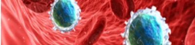
Stains and Dyes
Discover our range of stains, dyes, and reagents to visualize biological structures, check the pH of a sample, or identify the presence of contaminants in a solution. Our stains and dyes can be used in a multitude of applications, including for microbiology, histology, and protein staining.

Useful Links
Save Now - Exclusive Deals

Product Code 10407650

Product Code 10468420

Product Code 11472502

Product Code 11425392

Product Code 4530155

Product Code 4530134

Product Code 15634110

Product Code 15645620

Product Code 15688360
Must Have
Product Code 15614310

Product Code 7111838

Product Code 10483750

Product Code 18085552

Product Code 4532403

Product Code 11465501

Product Code 15654090

Product Code 11449911
Complete Your Order - Great Deals


FAQ
In histology, the terms "dye" and "stain" are often used interchangeably, but they have distinct meanings and applications:
Dye
A dye is a colored substance that can impart color to a material by binding to it. Dyes are typically soluble in water or other solvents and can be used to color various substances. In histology, dyes are the chemical compounds used to prepare staining solutions. They are the active ingredients that provide the color. Common dyes used in histology include hematoxylin, eosin, and methylene blue.
Stain
A stain is a solution or mixture containing one or more dyes that is used to color tissues, cells, or microorganisms to enhance visibility under a microscope. Stains often include additional components such as fixatives, mordants, or solvents to assist in the staining process. In histology, staining is the process of applying a stain to a biological specimen to highlight specific structures or components. Different stains are used to selectively color various parts of the tissue or cells. Common stains include hematoxylin and Eosin (H&E), gram stain, and periodic acid-SCHIFF (PAS) stain.
In summary dye refers to the chemical substance that provides the color. Stain refers to the prepared solution or mixture used to apply the dye to the specimen to visualize specific structures.
Several dyes are commonly used to stain bacteria, each serving different purposes depending on the staining technique and the type of bacteria being studied. Here are some of the most widely used dyes:
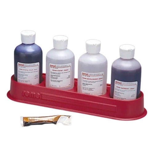
- Crystal Violet: Crystal violet is used as the primary stain in gram staining, which differentiates Gram-positive and Gram-negative bacteria. After applying crystal violet, gram-positive bacteria retain the dye and appear purple, while gram-negative bacteria do not retain the dye after the decolorization step
- Safranin: Safranin is used as a counterstain in gram staining. After the decolorization step, gram-negative bacteria are stained red or pink by safranin, while gram-positive bacteria remain purple
- Methylene Blue: Methylene blue is used in simple staining techniques and as a counterstain in various differential staining methods. It helps to visualize the shape and arrangement of bacteria
- Carbol Fuchsin: Carbol fuchsin is used in the Ziehl-Neelsen staining technique to identify acid-fast bacteria such as Mycobacterium tuberculosis. Acid-fast bacteria retain the red color of carbol fuchsin even after an acid-alcohol decolorization step, while non-acid-fast bacteria do not
- Malachite Green: Malachite green is used in spore staining techniques, such as the Schaeffer-Fulton method, to visualize bacterial endospores. Endospores retain the green color of malachite green, while the rest of the bacterial cells are counterstained with safranin
- Nigrosin or India Ink: Nigrosin and India ink are used in negative staining techniques to provide a dark background, making the transparent bacterial cells stand out. These dyes do not penetrate bacterial cells but stain the background, allowing visualization of cell shape and external structures
These dyes and staining techniques are fundamental tools in microbiology for identifying, classifying, and studying bacterial morphology and characteristics.
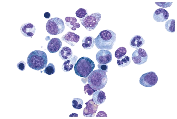
In histology, staining techniques are crucial for differentiating and visualizing various structures within biological tissues. The four main types of stains used in histology are:
1. Routine Stains
Hematoxylin and Eosin (H&E): This is the most commonly used stain in histology. Hematoxylin stains cell nuclei blue or purple, while eosin stains the cytoplasm and extracellular matrix pink or red. H&E staining provides a good overall view of tissue architecture and is used for general tissue examination.
2. Special Stains
These stains are used to highlight specific tissue components, structures, or microorganisms that are not easily visualized with routine stains. Examples include:
- Periodic Acid-Schiff (PAS): Stains carbohydrates and carbohydrate-rich structures, such as glycogen, mucin, and basement membranes, magenta
- Masson's Trichrome: Differentiates between collagen (blue or green), muscle (red), and cytoplasm (red)
- Silver Stains: Used to visualize reticular fibers, basement membranes, and certain microorganisms. Examples include Gomori's methenamine silver (GMS) stain for fungi and Warthin-Starry stain for spirochetes
3. Immunohistochemical Stains (IHC)
IHC stains involve the use of antibodies to detect specific antigens (proteins) in tissues. These stains are highly specific and are used to identify the presence and distribution of specific proteins, which can aid in diagnosis and research. Commonly used markers include Cytokeratins for epithelial cells, CD markers for different types of immune cells and Hormone Receptors such as estrogen or progesterone receptors in breast cancer.
4. Histochemical Stains
These stains are used to detect specific chemical components within tissues, often through enzyme reactions or other chemical processes. Examples include:
- Alcian Blue which stains acidic mucopolysaccharides and glycosaminoglycans blue
- Sudan Stains which stain lipids and fat cells. Sudan III, Sudan IV, and Oil Red O are commonly used for staining lipids red
- Prussian Blue which detects iron deposits, staining them blue